Optical Imaging of Tissue Mechanics via Laser Speckle Rheology
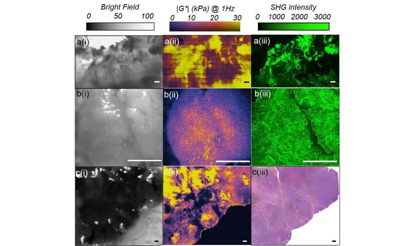
Bright-field images, LSR maps, and SHG images of human tissue specimens associated with different cancers. Credit: Figure 6 from DOI: 10.1117/1.JBO.25.5.050801 / SPIE
A majority of disease conditions from cancer and atherosclerosis to neurodegenerative and orthopedic disorders are accompanied by changes in tissue stiffness. Clinical medicine has long relied on manual palpation of suspected regions to detect tissue stiffness for diagnosis. Imaging modalities such as ultrasound, MRI and OCT can also effectively measure tissue stiffness. Laser speckle rheology (LSR) offers a new, noncontact optical approach. LSR uses an inexpensive laser similar to a common laser pointer to shine laser light on tissue—plus a camera to image speckle patterns reflected from light-scattering particles in the sample.Harvard Medical School associate professor of dermatology Seemantini Nadkarni, whose lab at the Wellman Center for Photomedicine developed LSR, explains the role of laser speckle patterns in communicating tissuestiffness: "In a soft, compliant specimen, light-scattering particles move about rapidly, causing the speckle pattern to fluctuate at a faster rate. In contrast, in a rigid specimen, a dense network of fibers restricts these motions, giving rise to a sluggish twinkling of speckle spots." By measuring the rate of speckle fluctuations, LSR enables measurement of the complex material properties of tissues.
Laser speckle rheology, a tutorial
A tutorial recently published by Nadkarni and her colleague Zeinab Hajjarian in the open access, peer-reviewed Journal of Biomedical Optics (JBO), provides a timely and visually rich review of optical methods in tissue mechanics, with a specific focus on LSR. The tutorial discusses various clinical applications for translating the LSR platform for applications in basic research and clinical medicine. It is based on a lecture series presented by Nadkarni at the 9th International Graduate Summer School, Biophotonics '19, in Hven, Sweden.
JBO Editor-in-Chief Brian Pogue, MacLean Professor of Engineering Science at Dartmouth College Thayer School of Engineering, says, "The tutorial combines the critical aspects of experimental methods in how speckle field measurements are designed and used, with an appreciation of the needs in cell, matrix, and biological tissue characterization." The authors review both in vitro applications and in vivo tissue imaging prior to summarizing the history of the field and where it appears to be headed.
With the goal of extending the reach of the LSR platform to the investigation and diagnosis of human disease from a biomechanical perspective, the research team has developed various devices that leverage the LSR platform for applications in hematology, interventional cardiology, and cancer research.
Palm-sized blood coagulation sensor
A major theme of investigation involves the use of LSR to detect bleeding and thrombotic disorders in patients at the point of care. The team has developed a palm-sized blood coagulation sensor, termed iCoagLab, that uses a few drops of blood to measure various parameters to evaluate a patient's coagulation state within minutes.
"Coagulation impairments happen when blood clots much too slowly or much too quickly, and the resulting clot is very loose or very firm causing bleeding or thrombotic events," said Nadkarni. "By measuring and monitoring the stiffness of clotting blood using LSR, we can identify life-threatening clotting defects and likely guide blood transfusion strategies in bleeding patients or inform dosing of blood thinners in patients with thrombotic conditions."
Mapping tissue stiffness within blood vessels
LSR can also be performed via small-diameter optical fiber bundles incorporated within endoscopes, catheters or needles to evaluate internal tissues inside the body that may otherwise be inaccessible for traditional mechanical testing. For instance, myocardial infarction, the leading cause of death worldwide, occurs due to the rupture of mechanically weak plaques within the vessel wall.
"Our research team has developed intravascular LSR catheter technology to yield the stiffness map of entire vessel walls and detect mechanically unstable fatty plaques implicated in heart attacks. We have tested the LSR catheter technology in coronary arteries from human cadavers and in living animals models, and are advancing the technology toward preclinical and clinical settings," said Nadkarni.
New LSR microscope to aid in cancer research
Various fundamental mechano-biology studies over the years have emphasized the importance of micromechanical aspects of tissue and disease and called for new tools for measuring these properties. The authors of this tutorial have also developed a new LSR microscope that enables mapping of the stiffness of tissues and materials at length scales over a few tens of microns.
"The new LSR microscope scheme captures the stiffness distribution map over several centimeters of a tissue sample in just a few seconds. In comparison, similar measurements obtained with traditional microindentation techniques could take several hours," said co-author Hajjarian, assistant in electrical engineering at the Wellman Center for Photomedicine, and instructor in dermatology at Harvard Medical School. "We have demonstrated the unique strengths of this technology in microfabricated phantoms, in vitro models of cancer malignancies, and tumor specimens collected from patients."
The authors touch on the technical aspects of how these physical principles drive different processing aspects of the speckle frame series to quantify mechanical parameters important to the tissue nature or function. Pogue finds this scheme remarkable: "In particular, the authors' approach to developing a flow chart of the processing algorithms and theoretical underpinnings provides the basics needed to understand what is measured."
By presenting LSR in the broader context of competing optical techniques and providing a comparative discussion of the attributes specific to each technology, the authors have served the interests of the biomedical optical engineering community. According to Pogue, "Their dissection of the competing optical methods used, such as optical coherence elastography, traction force microscopy, dynamic light scattering and diffusion wave spectroscopy helps put the methods of laser speckle rheology into better context, in the armamentarium of tools."
(From:https://phys.org/news/2020-05-optical-imaging-tissue-mechanics-laser.html)