Optical Coherence Tomography Shines With a New Type of Contrast
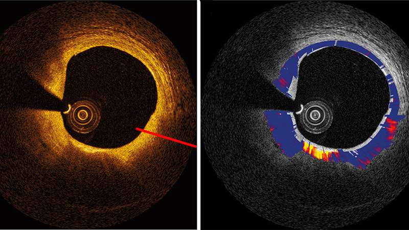
From Fig. 3, DOI: 10.1117/1.JBO.25.4.040901 Credit: SPIE
A diagnostic workhorse, optical coherence tomography (OCT), provides high-resolution 3-D imaging of subsurface tissue structures. Most likely, for your last eye exam, the ophthalmologist took cross-sectional pictures of your dilated eyes to measure the thickness of your retinal layers using OCT.Minimally invasive microscopic OCT imaging is widely affordable and recognized as a tool of choice. Minted in 1991 by Fujimoto's group at MIT, which first conjoined the elements comprising OCT, the technique has been evolving as a multimodal platform for decades as researchers have developed multiple ways to add functionality to OCT. One of the most promising to emerge is imaging OCT signal attenuation in tissue.
Like an echo composed of light
Based on the backscattered signal produced when an optical beam encounters living tissue, OCT enables researchers and clinicians to quantify structural thickening associated with various pathologies—from age-related macular degeneration to certain forms of cancer growth. But there are some ultra-small-scale changes in tissue morphology during the onset and progression of diseases beyond the scope of conventional clinically available imaging techniques. To capture such subtle yet significant changes, an important complement to standard OCT is quantitative measurement of the attenuation of the OCT signal. Signal attenuation can convey important information about the strength of the backscattered signal—like an echo composed of light, as it fades, it provides valuable information about the optical properties of the biological tissue.
Coming of age: standardize for greater reliability
OCT imaging of signal attenuation is the subject of a recent review by a group of senior OCT experts in the open access Journal of Biomedical Optics. The review synthesizes the state of the art, tracing its 15-year development. The authors tabulate values for a range of health applications—dermatology, ophthalmology, cardiology, and oncology—and provide a comprehensive vision of the scope of what is possible, where things are working, and where there is concern.
As the field continues to emerge with considerable interest, modeling the connection between tissue structure, optical properties, and measured quantities remains a formidable challenge. For instance, it is necessary to locate homogeneous tissue regions for attenuation calculation, and that is dependent on the specific tissue morphology. When tissue has layered structures, the OCT signal strength varies. For example, normal skin comprises epidermis (low signal) and dermis (high signal), which are sorted out or "segmented" to confine the depth ranges for measuring signal attenuation. In such circumstances, calculating attenuation may demand revision of methods according to specific disease conditions and simply may not be feasible for all conditions.
As an emerging approach, there is some controversy around the accuracy of the attenuation values and diverse approaches to estimating signal attenuation in OCT. The use of different systems and methodologies, as well as occasional lack of rigor in sample preparation or approach, has resulted in wide spreads of reported values for OCT signal attenuation values. The authors recommend standardization for rigorous methodology in future research seeking to contribute to the field.
As OCT imaging of signal attenuation comes of age, standardization can increase the reliability of the parameter. A prior review from a group at Vanderbilt Universitymade a first step to facilitate further investigation for advancing the clinical usefulness of OCT. The recently published review collects perspectives of several international groups involved in OCT research—including groups from University of Western Australia, University of Amsterdam and related medical centers in The Netherlands, and University of Surrey, UK—seeding the common ground needed to foster reliable new contrast potential in OCT.
(From:https://phys.org/news/2020-04-optical-coherence-tomography-contrast.html)