Laser Scanning Microscope Add-on Improves Large Tissue Volume Imaging
A team of researchers at Tel Aviv University (Tel Aviv, Israel) has combined commercially available hardware and open-source software to improve rapid 2D and 3D imaging of the brainand other tissues. The work could help scientists to better understand brain dynamics and discover new treatments for health problems such as stroke, epilepsy, and dementia. The combination approach, called PySight, serves as an add-on for a laser scanning microscope.
Because it can image deep into tissue, multiphoton microscopy is often used to study the rapid activity patterns of neurons, blood vessels, and other cells at high resolution over time. This microscopy method uses laser pulses to excite fluorescent probes, eliciting the emission of photons, some of which are detected and used to form 2D and 3D images.
Trying to capture the full breadth of neuronal activity with multiphoton microscopy forces scientists to image faster. As a result, fewer and fewer photons become available to form images. The challenge then becomes how to get meaningful images under these dim conditions.
"To tackle this challenge, microscopists have used a detector-readout method called photon counting," says research team leader Pablo Blinder. "However, because its implementation required extensive electronics knowledge and custom components, photon counting has never been widely adopted. In addition, commercially available photon counting solutions were ill-suited to perform very fast imaging such as required for 3D imaging. PySight's easy installation procedure and its integration with state-of-the-art hardware eliminate such concerns."
In addition to advancing neural imaging research, PySight’s improved sensitivity could facilitate rapid intraoperative identification of malignant cells in human patients using multiphoton microscopy. PySight's novel approach for reconstructing 3D scenes could also improve performance of light detection and ranging (lidar). This could help lower the costs of self-driving cars that use lidar to map their surroundings.
PySight provides high spatiotemporal resolution while producing a data stream that scales with the number of detected photons, not the volume or area being imaged. "Conventional data acquisition hardware stores the brightness of every pixel or 3D voxel even when it is zero, as no photons were detected in that particular location," Blinder explains. "PySight, however, only stores the precise detection time of each photon. If no photons were detected, nothing gets written to disk. This allows researchers to conduct rapid imaging of large volumes over long sessions, without compromising spatial or temporal resolution."
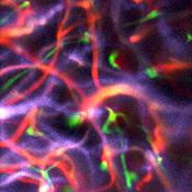
Brain vasculature in an anesthetized mouse captured using PySight is shown. (Image credit: Pablo Blinder, Tel Aviv University)
To reconstruct a multidimensional image, knowing when each photon hits the detector isn't enough. It's necessary to also know where it originated in the brain. "This is even trickier if you want to simplify the system and avoid synchronizing the different scanning elements," Blinder says. "To accomplish this, our software reads a list of photon arrival times along with timing signals from the scanning elements, determines the origin of each photon within the sample, and generates the corresponding 3D movies."
The photon arrival times are generated by a device known as a multiple-event time digitizer (multiscaler), which records the times with a precision of 100 ps. Another key component was an off-the-shelf resonant axial scanning lens that changes the focal plane hundreds of thousands of times per second. This lens was used to rapidly scan the laser beam across different depths within the brain and allowed the team to reconstruct continuous 3D images.
"The multiscaler we used hasn't been applied to neuroimaging because the output isn't easy to interpret, and using a resonant axial scanning lens for bioimaging has required custom-made scanning synchronization hardware or proprietary code to obtain the 3D data," Blinder says. “PySight turns the output from both components into a 3D movie effortlessly. As PySight is free and open-source software, it should greatly aid labs previously deterred by the high technical barrier that accompanied continuous 3D imaging." Further, having a generic application interface, PySight could also be used to interpret similar photon detection times from other suitable hardware devices.
To test whether PySight was truly plug and play, the researchers walked with their multiscaler to another imaging lab on the Tel Aviv University campus. They were able to simply plug in the device into the existing multiphoton microscope, download the PySight software, and start recording single-trail odor responses in fruit flies genetically modified to express voltage indicators. This fast probe for neuronal activity detects the finest aspects of neuronal activity yet it is considered too dim to be used without photon counting with this type of microscopy. PySight capabilities pave the road for and easy implementation of multiphoton voltage imaging in almost any laboratory.
In addition to continuing to improve the PySight software, the researchers would like to add support for other microscopy imaging methods such as fluorescence lifetime imaging (FLIM), which relies on the timing of each photon relative to its originating laser pulse. Because the software is open source and provides direct access to photon arrival times, it enables other scientists to add new features and meet their specific needs.
(From:https://www.bioopticsworld.com/articles/2018/09/laser-scanning-microscope-add-on-improves-large-tissue-volume-imaging.html)