Optical Imaging Methods Combine to Deliver Neural Tube Defect Images in Real Time
A neural tube defect develops in an embryo's neural tube—a hollow structure made of cells that eventually becomes the brain and spinal cord—prior to a woman learning that she is pregnant. Recognizing this, researchers at the University of Houston (UH; Houston, TX) and collaborators will use an optical imaging combination approach to delve into why the neural tube closes in most embryos but remains open in others, leading to birth defects such as spina bifida and anencephaly.
UH professor of biomedical engineering Kirill Larin, together with Baylor College of Medicine (also in Houston) professor Richard H. Finnell and Giuliano Scarcelli, assistant professor of bioengineering at the University of Maryland (College Park, MD), will combineBrillouin spectroscopy and optical coherence tomography (OCT) to deliver three-dimensional (3D) images of the mechanical factors at play when the neural tube closes and when it does not.
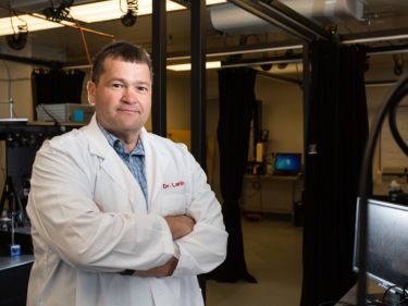
UH professor of biomedical engineering Kirill Larin tackles neural tube defects by utilizing new technology to peer inside embryos.
Most commonly used to examine the retina, high-resolution OCT uses light waves to take cross-section pictures. Brillouin spectroscopy is a light-scattering technology that will sense the stiffness of the tissue, which is critical to its success in closing. During malfunctions there are areas of low stiffness, so it is not folding together properly, Larin says.
"We will create this hybrid microscope putting these two powerful technologies together," Larin explains. "OCT will image the development of the neural tube while at the same time, Brillouin spectroscopy will probe its mechanical properties. We will be imaging and sensing at the same time."
Finding out how mechanical properties controlling neural tube closure in developing embryos can be manipulated to ensure proper neural development is the first step to exploring treatment for the defect. "If we find out what causes the tube to close, what is exactly happening, we can develop new drug treatments for at-risk embryos," Larin says.
The work is receiving $3.2 million in funding from the National Institutes of Health (NIH).