OCT Imaging Technique Could Improve the Diagnosis, Treatment of Glaucoma
Seeking to advance glaucoma diagnosis and treatment, researchers at the Indiana University School of Optometry (Bloomington, IN) have developed a noninvasive ophthalmoscopy method to visualize and count individual cells at the back of the eye.
Related: Optical device uses adaptive optics to detect diabetic retinopathy early
Zhuolin Liu, the first author on the paper describing the work and a research associate in Donald T. Miller's laboratory at the time of the study, says the new method is an optical microscope with extremely high resolution and sensitivity. It enables eye care practitioners to directly count retinal ganglion cells (the cells that die in glaucoma), allowing them to assess glaucoma damage long before current clinical instruments allow them to, she says.
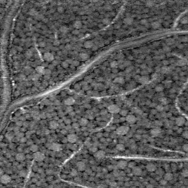
Donald T. Miller's ophthalmoscopy invention shows tightly packed retinal ganglion neurons that are disrupted only by blood vessels and capillaries.
Glaucoma causes blindness by damaging cells in the retina, which transforms light into electrical impulses and transmits them to the brain. Miller, a professor in the Indiana University School of Optometry, says that thousands of cells must die before current clinical methods detect glaucoma.
"The cells are very small, highly translucent, and only dimly reflective. This prevents clinical instruments from creating sufficiently sharp images to see the damage," Miller says. "If eye care practitioners could see this damage earlier, when it affects individual cells, it could dramatically improve the diagnosis and treatment of glaucoma and other diseases of the retina."
The research team's method improves the performance of optical coherence tomography (OCT), which is the primary method used to detect eye disease in a clinic. Miller's laboratory obtains images that have higher resolution and higher contrast by using adaptive optics. The technology was originally developed in astronomy to remove the effects of atmospheric blur from telescope images.
"Our method provides a form of retinal biopsy," Liu says. "But it uses light, so it is noninvasive and allows repeated assessment of the same cells. By refocusing the instrument, we can obtain sharp images of cells at any depth in the retina, from cells on the surface to those that line the retinal bottom and keep the retina functioning."
Miller and his research associates are working to make the new method sensitive to the physiological activity of cells. "If we can achieve this, it opens new, previously unimagined ways to map functional aspects of retinal neural circuitry and to detect the earliest cellular changes associated with disease onset," he says.
Miller disclosed the new method to the IU Innovation and Commercialization Office (ICO), which protects, markets, and licenses intellectual property developed at Indiana University so it can be commercialized by industry. IU ICO has applied for a patent on the method.
"Integrating the method into a commercial OCT product for eye care will require federal approval," Miller says. "A commercial version could be readily made much smaller than the current laboratory version to meet requirements for clinic use."
(From:https://www.bioopticsworld.com/articles/2018/03/oct-imaging-technique-could-improve-the-diagnosis-treatment-of-glaucoma.html)