Modulating Speckle for Better OCT Imaging
Optical coherence tomography (OCT) has become invaluable to medical imaging over the past quarter century—the same length of time that scientists have been wrestling with the technique's inherent speckle noise. Now a team at Stanford University, USA, has tweaked OCT technology to reduce speckle noise and reveal fine-structure details within living tissues (Nat. Commun., doi: 10.1038/ncomms15845).
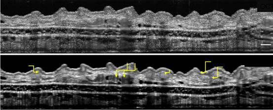
Top: Conventional OCT imaging of a mouse’s outer ear (pinna). Bottom: SM-OCT image of the same pinna reveals additional details, highlighted with yellow arrows. [Image: de la Zerda Lab, Stanford University]
Attacking noise
As all physics students learn in their first laser experiments, speckles inevitably arise from the mutual interference of coherent wavefronts. Commercial OCT systems fight the resulting image degradation in several ways: incoherently averaging multiple images, for example, or using image-processing techniques that mask the appearance of speckle noise without revealing lost details.
Scientists in the research group of Stanford structural-biology professor Adam de la Zerda call their technique speckle-modulating OCT (SM-OCT). Conceptually, the scheme introduces time-varying local phase shifts within the light beam that illuminates the sample tissue. The reflected light within the same voxel also has local phase shifts. Over time, an unlimited number of uncorrelated speckle patterns can be incoherently averaged to produce images with virtually no speckle noise.
Off-the-shelf systems
SM-OCT requires no new medical devices; the team created their prototypes out of two different commercially available OCT systems, plus a ground-glass diffuser and various lenses and other pieces of optical-bench equipment. The diffuser, placed in the line of sight between lenses, moves to a slightly different position between each round of imaging scans. The averaged scans showed much better resolution that conventional OCT. The group tested its prototype devices by imaging the retina and ear pinna of an anesthetized mouse as well as the fingertip ridges of a human volunteer.
According to lead author Orly Liba, a Stanford doctoral candidate in electrical engineering, the team's biggest experimental challenge was implementing the local phase changes onto the tissue sample. Simply placing the moving diffuser on top of the sample did not make the imaged region and the local phase variations overlap optically. “We solved this issue by duplicating the image plane using two lenses in a 4f configuration,” she says. “This way the diffuser effectively (optically) coincides with the imaged region in the sample.”
Toward clinical testing
Next, the researchers want to take SM-OCT into the clinic and test its capabilities on human retinal imaging, skin-cancer diagnosis and intra-operative detection of tumor margins. “We believe that SM-OCT will reveal structures with a resolution equivalent to a microscope (but without needing to slice the tissue) and that it will enable better diagnostic and operative capabilities (compared to conventional OCT),” Liba says.
OSA Honorary Member Steven Chu was one of the co-authors on the study.
(From:https://www.osa-opn.org/home/newsroom/2017/june/modulating_speckle_for_better_oct_imaging/)